 Scgf
Scgf gene is unexceptionally more or less up-regulated in most leukemia cells regardless of acute or chronic leukemia (
18,
142,
143,
149,
Pat21-
Pat24,Pat27), leukemia cell lines (
6,
171-
174) and lymphoma (
148,
Pat21,Pat27) or myeloma cells (
Pat21,Pat27). Leukemia occupies most of top 10 cancers in
scgf gene expression (
Genevisible). The
scgf up-regulation is more prominent in drug-resistant leukemia (
142,
147) and early relapse in acute leukemia (
144). Asparaginase-resistant genes such as IGFBP7 but not
scgf are up-regulated in pediatric B-lineage ALL cells cocultured with allogeneic BM stromal cells (
370).
Scgf gene expression is significantly down-regulated in the CD10
+ leukemia cells from pediatric precursor B-ALL patients with a better prognosis compared to other CD10
- ALL cells (
402).
Scgf gene expression is up-regulated in non-Down syndrome acute megakaryoblastic leukemia (AMKL) cells with
CBFA2T3-GLIS2 fusion gene compared to non-Down syndrome AMKL cells with other fusion genes, e.g.
OTT-MAL(218).
As described above,
scgf up-regulation is estimated a second hit to induce overt leukemia following a first hit of mutated
N-rasG13R (39),
MLL/FOXO3A in secondary ALL (
145),
AML1/MTG16 in AML t(8:21)(q22:q22) and AML t(16:21)(q24:q22) (
146), inv(16), mutated RUNX1-T-7 and NPM1/NRAS in AML (
597),
PML/RARΑ in APL t(15:17)(q22:q21) (
222,543),
PLZF/RARΑ in APL t(11:17)(q23:q21) (
222,543),
MLL translocation in t(4:11) biphenotypic B-AML and ALL (
169), 5q- abnormality (
170),
E2A/PBX1 in t(1:19) B-ALL (
Pat20) and recombinant
MLL in B-ALL (
Pat20). BM plasma SCGF level is highly elevated and Flt3L declined in NPM1-mutated
de novo AML compared to other AML subtypes (
711).
Scgf gene expression is relatively up-regulated in mixed-lineage leukemia (MLL) cells compared with ALL cells (
316), particularly in AML with a
MLL translocation but not in AML with a partial tandem duplication of
MLL (
327). Highly up-regulated
scgf in a single pediatric AML blast is correlated with poor overall survival compared to low
scgf expression (
694). In contrast,
scgf gene expression in high-risk elderly AML is significantly down-regulated compared to low-risk AML (
586). AML with high
scgf expression is associated with prolonged survival compared to AML with low
scgf expression (
650,706).
Scgf highly up-regulated AML is associated with better prognosis than modestly up-regulated AML (
641), and AML with hypermethylated
scgf DNA is poorly induced to remission compared to
scgf-hypomethylated AML.
As for pediatric AML,
scgf is up-regulated and its promoter region is demethylated in BM leukemia cells from MLL-rearranged IL-6
hi/IL-6R
hi pediatric AML patients with poor prognosis (
683). Total and north shore of
scgf promoter area is hypermethylated in M0 and M1 AML cells compared to M2-M5 AML cells and CpG island and south shore (
703).
Scgf methylation correlates with cytogenetic abnormalities but not with age, gender and race of AML patients.
Scgf gene expression is highly up-regulated in GPR84-expressing Lin
-Sca-1
+c-kit
+ pre-leukemic stem cells (LSCs) from MLL-AF9-transformed mice relative to vector controls (
477).
Scgf gene expression is up-regulated in t(8;21)-positive AML cells independent on
POU4F1 gene dysregulation (
247).
Scgf gene signature is observed in the AML cases without abnormalities, e.g.
flt3 TKD, 3q and del5(q) (
230).
In addition to the therapy hitherto for arespective entrance to leukemogenesis, blocking an exit common to leukemogenesis, i.e. SCGFmediated proliferation of leukemia cells, could be a potential master key therapy versatile for any type ofleukemia, since anti-SCGF antibody eradicates
in vitro growth of five types of leukemia cell line (
141). Anti-SCGF antibody-induced suppression is relatively late onset, indicating a possible action of SCGF on a certain subset of leukemia cell lines, e.g. Hoechst 33342-excluding side population equivalent to LSCs.
Scgf gene expression is up-regulated in CD34
+ AML cells containing LSC population compared to CD34
- AMLcells (
355). However,
scgf expression in LSC-equivalent CD34
+CD38
- AML cells is not different from that in CD34
+CD38
+ AML cells (
168). AML-derived CD34
- LSCs express
scgf gene, however it is down-regulated relative to normal myeloid progenitor cells (
Pat29).
Scgfr but not
scgf is predicted to be up-regulated in LSCs.
CpG sites of
scgf gene promoter are differentially hypomethylated in the pediatric ALL cells at relapse as compared to the matched cells at diagnosis (
344). DNA methylation is lost at
scgf CpG sites in pediatric B-ALL cells without
BCR/ABL1 and
MLL translocations compared to normal B cells (
404).
Scgf gene is differentially expressed and methylated in pediatric ALL cells compared to normal CD19
+ cells (
617).
Higher
meningioma 1 gene expression in the BM or PB cells from patients with untreated cytogenetically normal AML negatively correlates with
scgf gene expression (
207).
WEB-2170 (an inverse agonist of platelet-activating factor receptor)-induced apoptotic human APL NB4 cells down-regulate
scgf gene expression as compared with untreated controls (
213).
Direct target genes for PLZF-RARΑ (promyelocytic leukemia zinc finger-retinoic acid receptor Α) fusion protein as seen in the translocation t(11:17)(q23:q21) of acute promyelocytic leukemia (APL) cells were investigated using chromatin immunoprecipitation promoter and U133 Plus 2.0 expression arrays for U937T:PLZF-RARΑ-inducible cell line (
222). Bioinformatic analysis using PANTHER, Ingenuity Pathway Analysis indicates that PLZF-RARΑ antagonizes PLZF-mediated repression of
c-myc promoter to induce
c-myc and ontologically up-regulates
scgf gene in APL relative to other AMLs.
Scgf gene expression is up-regulated in plasmacytoid DC leukemia cells compared to normal plasmacytoid DCs (
642).
Scgf gene expression is significantly down-regulated in the bone marrow MSCs from patients with refractory anemia with excess blasts, and in the normal MSCs cultured with TGF-β1 (
588).
Scgf gene expression is down-regulated in bone marrow CD34
+ cells of CML patients relative to healthy controls (
296).
UBE2A (ubiquitin-conjugating enzyme E2A) mutation is specifically demonstrated in leukemia cells from patients with CML in blast crisis (
606).
UBE2A silencing with shRNA significantly up-regulates
scgf gene expression in CML blast crisis-derived K562 and 32Dcl3 cells.
CpG island of
scgf gene is hypermethylated in CLL cells without immunoglobulin heavy chain variable region mutation compared to normal mature naive B cells (
392). SCGF is well associated with β2 microglobulin as a prognostic marker in CLL patients treated with chemo-immunotherapy (
629).
Macrophages induced from U937 cells with phorbol myristate acetate express
scgf gene (
271).
SCGF is produced and secreted by human T lymphoma cell line Jurkat (
255, 460).
Scgf gene expression is down-regulated in the Jurkat cells transfected with HIV-1 Tat first exon encoding 1-72aa, but not in the cells with full-length Tat encoding 1-101aa (
262).
One of DNA copy number gains maps to chromosome 19q13.33 including
scgf gene in CD30
+ Hodgkin and Reed-Sternberg cells laser-capture microdissected from Hodgkin lymphoma samples (
323). Serum SCGF level shows no significant difference between HIV
+ B-cell non-Hodgkin lymphoma and HIV
+ controls without any malignancy (
463). NF-κB activation as seen in API2-MALT1
+ gastric MALT lymphoma is linked to down-regulation of
scgf gene at chromosome 19q13.2-q13.4 (
232). Causal regulator active in the tumor tissue from patients with diffuse large B cell lymphoma (DLBCL), TNFSF11, up-regulates
scgf gene expression (
383).
Scgf gene expression is down-regulated in CD5
+ DLBCL tissues as compared to CD5
- DLBCL tissues (
500).
Scgf gene expression is down-regulated in the bone marrow CD34
+CD38
low cells from patients with relapsing lymphoma or multiple myeloma one year after undergoing autologous peripheral blood stem cell transplantation compared to normal bone marrow controls (
437).
Bone marrow fibroblasts (BMFs) secrete elevated levels of IL-6, CXCL5 and M-CSF at the stage of monoclonal gammopathy of undetermined significance (MGUS), IGF-II at the stage of MGUS and multiple myeloma (MM), and SCGF at the stage of MM, as compared with normal BMFs (
206), suggesting that they can be a therapeutic target for preventing tumor progression.
Scgf gene promoter is hypomethylated in the bone marrow CD138
+ plasma cells from patients with MGUS compared to early stage of MM and healthy controls (
407).
Scgf gene overexpression in 24 IL-6-dependent and16 IL-6-independent myeloma cell lines significantly correlates with poor overall and event-free survival, indicating that
scgf gene overexpression can be one of the bad prognostic indices for MM (
312,Pat26).
Scgf is ranked the 6th critical gene signature shared among seven gene expression profiling datasets of MM (
349). Myeloma cells with t(4;14)(p16.3;q32) overexpress gene network between
MMSET/WHSC1,
FGFR3 and
scgf (365). Weighed gene co-expression network analysis discloses
scgf as a hub gene in multiple myeloma cells.
Scgf gene expression is highly up-regulated in t(4;14)-positive myeloma cells with
MMSET overexpression. Knockdown of t(4;14)-positive KMS-26 and NCI-H929 myeloma cells with
scgf siRNA down-regulates expression of
NFkB and
IRF4, a target for
MMSET, and decreases cell viability by 60% associated with increased annexin V-positive apoptotic cells. The findings indicate that
scgf can be a potential therapeutic target for t(4;14)-positive myeloma (
536).
No mutations in
scgf gene are detected in the granulocytes from patients with myeloproliferative disorder including
polycythemia vera, essential thrombocythemia and primary myelofibrosis (
452).
Genetic predisposition to overall clonal hematopoiesis is correlated with elevated circulating SCGF level (
679).
Other hematological disease
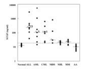
An SCGF concentration in the serum is an optimal index for monitoring hematopoietic recovery following stem cell transplantation (SCT) (
34). Plasma SCGF level falls down to one tenth of the preconditioning level after conditioning for allogeneic SCT, and gradually rises through hematopoietic recovery (
373). An increase in the serum SCGF level parallels hematopoietic recovery after SCT, and is not affected by GVHD, concomitant infection, blood transfusion and G-CSF administration. SCGF begins to rise earlier in autologous SCT and PBSCT than in allogeneic SCT and BMT, respectively. SCGF is superior to other indices in predicting hematopoietic recovery after SCT because SCGF can be easily measured by ELISA, serum is easily obtainable as compared with RNA, and ELISA can be done rapidly within several hours.
Mouse SCGF restores hematopoietic defect in CDH5-MAPK mice and irradiation-induced myelosuppression in wild type mice. Selective MAPK-activation in bone marrow endothelial cells from CDH5-MAPK mice up-regulates MAPK-downstream NFkB to induce perivascular niche inflammation (
616,Pat34). Disruption of endothelial cells leads to profound hematopoietic defect including exhaustion of hematopoietic stem (HSCs)/progenitor cells, decreased quiescent HSCs, elevated HSC apoptosis, decreased competitive repopulating ability of HSCs and decreased peripheral blood cells with myeloid-biased output. Plasma SCGF level is dramatically down-regulated in CDH5-MAPK mice, which is normalized by NFkB inhibition. Mouse SCGF resolves hematopoietic and vascular defects when administered to CDH5-MAPK mice or irradiated wild type mice.
Scgf gene expression is down-regulated in the CD34
+ bone marrow cells from children with very severe aplastic anemia (
389).
Hematological toxins influence on SCGF. Suppressionof SCGF production by malarial pigment hemozoin (PfHz)-phagocytized monocytes is one of the causes underlying childhood severe malarial anemia (
175). Malarial pigment, PfHz (
Plasmodium falciparum Hemozoin), is phagocytized by monocytes to suppress their cytokine production.
Scgf is the gene most differentially expressed in the normal PB-MNCs cultured with PfHz, i.e.
scgf gene is down-regulated to the level of <40% relative to the untreated control. The finding is confirmed by RT-PCR. SCGF level in the plasma and cultural supernatants of PB-MNCs from children with severe malarial anemia (SMA, Hb<6.0g/dl) is significantly lower than normal controls, the former of which positively and negatively correlates with Hb level and reticulocyte production index (RPI), and the percentage of PfHz-containing monocytes, respectively. Plasma SCGF level in the case with RPI<2 is significantly lower than that with RPI>3. Mechanism underlying childhood SMA is thought to be multifactorial, and one of the important causes is suppression of SCGF production by PfHz-phagocytized monocytes. To further clarify a functional role of SCGF in SMA, it was investigated whether homozygous T allele in the
scgf promoter (SNP
[c/t]rs7246355) at the position of 539 nucleotides upstream from the first exon was associated with the development of SMA in children with
P. falciparum malaria.
Scgf genotyping indicated a significant prevalence of TT allele in non-SMA (6.0g/dl≤Hb<11.0g/dl) and CC allele in SMA. SCGF levels in the plasma and cultural supernatants of PB-MNCs from
P. falciparum-infected children with TT allele were higher than those with CC allele. In addition, TT allele groups had high reticulocyte production index relative to CC allele groups. Consequently
scgf promoter variant (SNP
[c/t]rs7246355) protects against the development of childhood SMA (
221).
Concomitant infection little affects SCGF production, e.g. SCGF level in the serum from patients infected with evolavirus (
272) or dengue virus 2 (
295) does not differ from healthy controls. Plasma SCGF level appears a little higher than controls on day 2 and 3 of acute phase of Chikungunya viral infection (
325).
SCGF production is highly repressed in human macrophages infected
in vitro with influenza viruses, particularly seasonal H1N1 and H3N2 viruses (
322). However plasma SCGF level is significantly elevated in patients with H7N9 influenza A viral infection as compared to normal controls, and can be one of prognostic biomarkers to predict fatal outcome (
502).
Another report is apoptosis induced by toxic metal, cadmium;
scgf gene is up-regulated when CCRF-CEM cells are incubated with 20μM cadmium for 6 hours (
176).
Infection
Serum SCGF level determines onset of SARS-CoV-2 infection. Machine learning indicates with 100% accuracy that SARS-CoV-2-infected patients with higher (>127ng/ml) and lower (<127ng/ml) serum SCGF level are asymptomatic and symptomatic respectively (
632). When SCGF is excluded, serum levels of IL-16 (>45pg/ml) and M-CSF (>57pg/ml) are asymptomatic marker. SCGF is a prognostic biomarker for symptom onset and, more importantly, SCGF could control a novel protective immune mechanisms against infection. Plasma SCGF level is up-regulated in patients with SARS-CoV-2 pneumonia (
651). Plasma SCGF level is higher in COVID-19 patients than healthy controls (
654, 655), and the elevated level is significantly lower in severe COVID-19 patients with pre-existing asthma than those without asthma (
654). Higher plasma SCGF level is associated with endothelial disruption in severe SARS-CoV-2 infection (
661). Plasma SCGF level is significantly lower in COVID-19 convalescent donors with high expression of PLAU, IL-1B, NFKB, PLEK and LCP2 compared to healthy controls (
684). As for 2018-2019 outbreak of Andes orthohantavirus pulmonary syndrome in Argentina, reduced serum SCGF level is associated with hantavirus spreaders compared to non-spreaders, and among spreaders, notably with superspreaders compared to non-superspreaders (
636). Like SARS-CoV-2 infection SCGF could be a critical player of innate immunity against hantavirus infection.
Scgf gene expression is up-regulated in human peripheral blood mononuclear cells 4hrs after
in vitro Rhizopus oryzae infection and down-regulated 24hrs afterwards (
640).
Elevated plasma SCGF level before HIV infection correlates with increased viral load and decreased CD4
+ T cells after infection (
670).
SCGF level is down-regulated with all other factors elevated in the cerebrospinal fluid from pediatric patients with enterovirus encephalitis (
673).
Scgf gene expression is up-regulatedin vitro in rainbow trout head kidney cells in response to fungal PAMPs
including β-glucan peptide and furfurman (663). SCGF could exhibit innate immunity against fungus infection, e.g.
Malassezia furfur, like same C-type lectin dectin-2 that is a receptor for furfurman.
Oncology
Here described are SCGF-related findings in general oncology. Also see malignancy and disease of
brain,
eyes,
bone,
cardiovascular system,
lung,
breast,
liver,
kidney, gastrointestinal tract, prostate,testis,ovary,uterus,placenta and
skin,
c
Visit
The Human Protein Atlas where are presented a bonanza of histochemical staining data in cancer tissues.
When ovarian cancer tissue is cultured with chimeric single chain variable fragment (scFv) anti-SCGF antibody, tumor-associated macrophages are modulated to recruit TILs, then they induce apoptosis in ovarian cancer cells without affecting normal cells (
Pat37). Apoptotic effect of the antibody is demonstrated in other solid tumor cells. The antibody further synergizes with Nivolumab to promote apoptosis of Nivolumab-resistant cancer cells. The findings indicate that inhibition of SCGF opens the way to a new cancer therapeutic regimen.
Scgf gene expression in cancer tissues usually indicates unfavorable prognosis in colorectal, lung, renal, testicular, thyroid and urothelial cancers (
562). A higher plasma SCGF level is a biomarker signature predicting neoplasm-related 10-year mortality (
599).
When metastatic carcinoma cells and surrounding stromal cells are analyzed on their specific gene expression by the method of laser capture dissection,
scgf gene is significantly up-regulated in the stromal cells (
150). The findings indicate that surrounding tissue excessively produces SCGF to accelerate an overgrowth and extension of the carcinoma cells into the circumferential milieu. SCGF is the most important cytokine capable of reinforcing metastasis, tumor formation and self-renewal of cancer cells, essentially cancer stem cells. Anti-cancer drug surviving cells (DSC) are positive for CD133, SSEA-3 and Oct4, and may well be virtual cancer stem cells with self-renewal and tumor-forming ability in SCID mice (
151). An SCGF concentration in the cell lysate and conditioned medium of DSCs is strikingly higher than that of drug-unsorted original cancer cells. The tumor-promoting activity of SCGF can be one of the targets for tumor suppressor.
Scgf gene is down-regulated in the cancer cells transfected with tumor suppressor gene, semaphorin 3B (
177) or p53 (
116). Consequently stem cell-specific SCGF and/or SCGFR is a potential target for cancer therapy (
116,141,
150,
151,
177).
Extracts of NCCIT cancer cells dedifferentiate and reprogram 293 normal epidermal cells, and up-regulate
scgf gene 2-6 weeks before Oct4, Nanog, Sox2 and Oct4-responsive genes of UTF1 and REX1 (
159,Pat25). The 293 cells exhibit stem cell nature capable of differentiating into neuronal, adipose, osseous and endothelial cell lineages.
When CD133 gene expression of human malignant melanoma FEMX-I cells is suppressed using CD133 nucleotide sequence 773-792-targeted shRNA, Wnt inhibitors and
scgf genes are up-regulated as compared with untreated controls (
162). CD133 is one of the markers of cancer stem cells. Down-regulation of CD133 reduces tumorigenic and metastatic ability of FEMX-I cells, consistent with up-regulation of Wnt inhibitors. Simultaneous
scgf up-regulation implicates some compensatory mechanisms for proliferative activity of cancer stem cells.
Mouse Krebs-2 ascites carcinoma cells internalizing dUTP-5'-TAMRA-labeled human 500bp
Alu repeat are tumor-initiating stem cells that specifically express "stemness"
scgf gene (
555).
SCGF interacts with splice variant 2 but not with splice variant 1 of methionine adenosyltransferase 2Β up-regulated in colon cancer cell line, RKO (
257), which could regulate transcription and give a growth advantage to cancer cells.
Scgf gene expression is down-regulated in the quadriceps from severely cachectic mouse transplanted with Colon26 carcinoma cells, in contrast to a marked up-regulation of IL-6 and STAT3 pathway genes (
333).
Scgf gene expression is differentially down-regulated in the gastrocnemius muscle from
KrasG12D/+Lkb1f/f mice with nonsmall cell lung cancer-associated cancer anorexia cachexia syndrome compared to fasted normal mice (
577).
Refer to
Oncomine where
scgf gene regulation in malignant tissues has been archived.
miRNA
Disease-related modulation of
scgf-targeting miRNA is described in
Gene miRNA section.